Streak Retinoscopy, a revolutionary technique pioneered by the Khanna Vision Institute, is transforming the way eye care professionals diagnose and manage refractive errors. This innovative approach, developed by the renowned ophthalmologists at the institute, offers a highly accurate and reliable method for measuring the eye’s focusing power, known as the refractive state.
Unlike traditional retinoscopy techniques, Streak Retinoscopy utilizes a specialized instrument that projects a narrow beam of light onto the patient’s eye. By observing the movement and behavior of this light streak, eye care professionals can precisely determine the eye’s refractive status, including the presence and degree of nearsightedness, farsightedness, and astigmatism.
The Khanna Vision Institute’s expertise in Streak Retinoscopy has set a new standard in the field of optometry and ophthalmology. The institute’s clinicians have undergone extensive training and have honed their skills to perfection, ensuring that patients receive the most accurate and reliable diagnoses possible.
One of the key advantages of Streak Retinoscopy is its ability to provide objective and quantifiable data, which is crucial for developing the most appropriate treatment plan. By accurately measuring the eye’s refractive state, eye care professionals can prescribe the precise corrective lenses or other interventions needed to restore clear and comfortable vision.
Equipment Needed
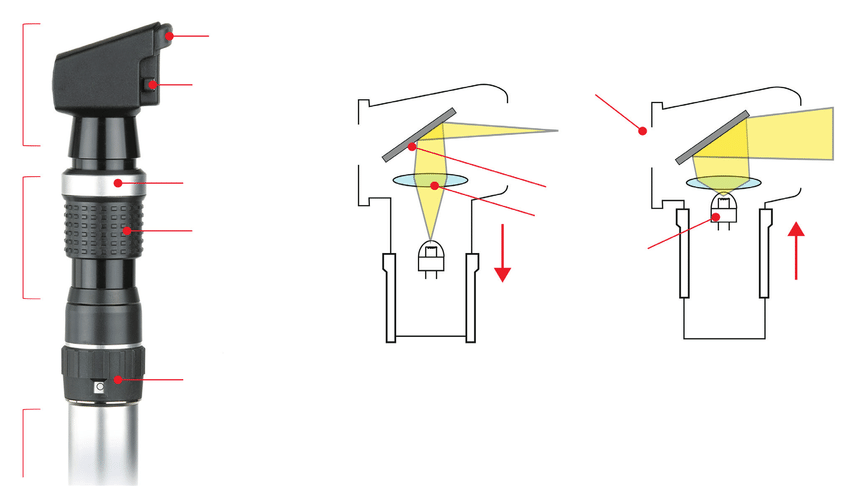
Retinoscope: A handheld device that projects a streak of light into the eye.
Trial Lens Set or Phoropter: Used to place lenses of varying powers in front of the patient’s eye to neutralize the reflex seen through the retinoscope.
Basic Principles
Retinoscope: The retinoscope projects a streak of light into the patient’s eye. This is done by moving the streak of light and observing the movement of the reflex (the light reflected from the retina), the examiner can determine the refractive error.
Reflex Movement: The movement of the reflex observed through the retinoscope helps the examiner understand whether the eye is myopic (nearsighted), hyperopic (farsighted), or astigmatic (having irregular curvature).
Procedure
Preparation:
The testing room should be dimly lit to ensure the retinoscope light is easily visible.
The patient should be seated comfortably, looking at a distant target to relax accommodation.
Initial Observation:
The examiner stands about one arm’s length from the patient and directs the streak of light into the patient’s eye.
The retinoscope is moved back and forth to observe the reflex movement.
Neutralizing the Reflex:
The examiner introduces lenses of different powers in front of the patient’s eye using a trial lens set or phoropter.
The goal is to find the lens power that stops the movement of the reflex (neutralization).
With Movement:
If the reflex moves in the same direction as the retinoscope streak, the eye is hyperopic (requires a converging lens).
Against Movement:
If the reflex moves in the opposite direction, the eye is myopic (requires a diverging lens).
Determining Refractive Error:
The power of the lens that neutralizes the reflex is noted.
For astigmatism, different meridians are examined by rotating the streak of light to different angles, and cylindrical lenses are used to correct the specific refractive error in each meridian.
Fine-Tuning:
The procedure is repeated for the other eye too.
Final adjustments are made to ensure the most accurate prescription.
Types of Reflex
With Movement:
Reflex moves in the same direction as the retinoscope streak, indicating hyperopia or less myopia than the working distance.
Against Movement:
Reflex moves in the opposite direction to the retinoscope streak, indicating myopia greater than the working distance.
Neutral Reflex:
No movement of the reflex, indicating the correct lens power has been found for that meridian.
Considerations
Working Distance:
The distance between the examiner and the patient must be considered when calculating the final prescription. A typical working distance is 67 cm (1.5 D).
Accommodation Control:
Ensuring the patient’s accommodation is relaxed is crucial for accurate measurements.
Advantages of Streak Retinoscopy
Provides an objective measurement of refractive error.
Useful for patients who cannot communicate their visual needs.
Can detect irregularities and asymmetries in refractive error (astigmatism).
Moreover, the Khanna Vision Institute’s commitment to innovation and continuous improvement has led to the development of advanced techniques and technologies that complement Streak Retinoscopy. These include the use of cutting-edge diagnostic equipment, such as wavefront analysis and corneal topography, to provide a comprehensive assessment of the eye’s structure and function.
In conclusion, the Khanna Vision Institute’s expertise in Streak Retinoscopy is a testament to its dedication to delivering the highest quality eye care. By leveraging this innovative technique, the institute’s clinicians are able to provide patients with accurate diagnoses and personalized treatment plans, ensuring optimal visual outcomes and improved quality of life.