Topography-Guided Photorefractive Keratectomy (PRK) is an advanced form of laser eye surgery used to correct refractive errors such as myopia (nearsightedness), hyperopia (farsightedness), and astigmatism. This advanced procedure utilizes detailed corneal mapping to precisely reshape the irregular corneal surface, effectively addressing the visual distortions associated with this condition.
The Khanna Vision Institute has established itself as a leader in the field, offering this innovative treatment option to patients. By leveraging the power of topography-guided technology, the skilled surgeons at the institute are able to create a customized treatment plan that targets the specific irregularities of each patient’s cornea.
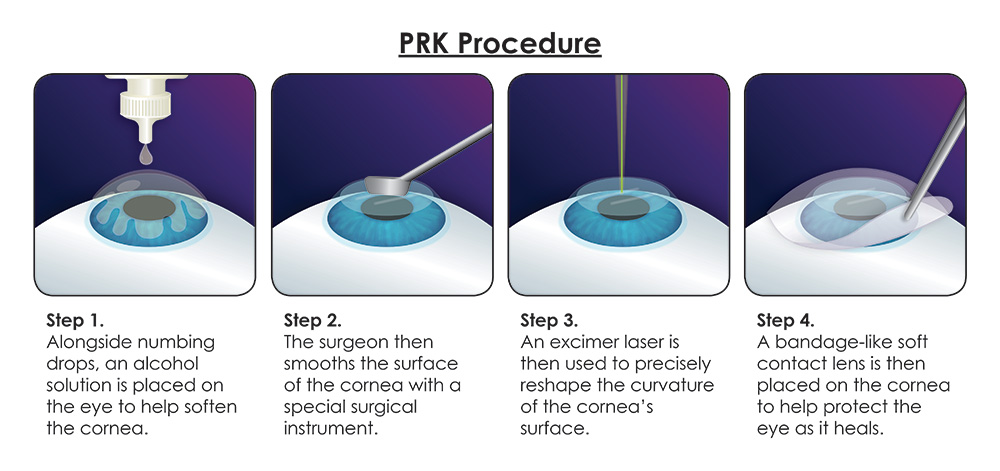
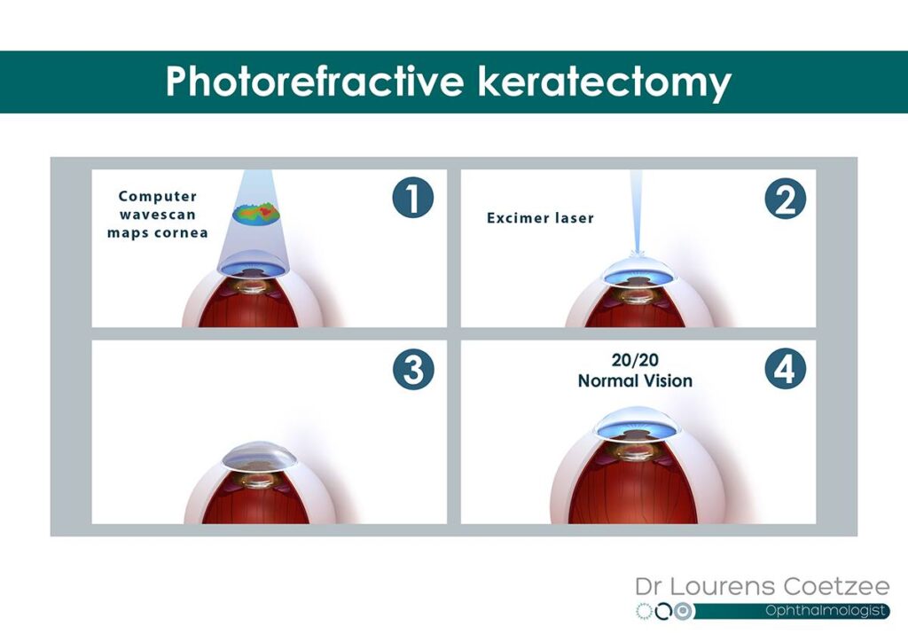
During the procedure, the surgeon removes the corneal epithelium, allowing direct access to the underlying stromal layer. Using the topography data, the laser is then programmed to selectively remove microscopic amounts of corneal tissue, gradually reshaping the cornea and improving its optical properties. This precision-based approach helps to minimize the risk of complications and ensures a more predictable visual outcome for the patient.
Key Points of Topography-Guided PRK
Customization:
Unlike traditional PRK, which uses a standard laser pattern, topography-guided PRK tailors the laser treatment to the unique shape and irregularities of the patient’s cornea. This results in a more personalized and potentially more effective correction.
Healing and Recovery:
The epithelium regenerates over a few days following the procedure. Patients may experience discomfort and blurred vision during this healing period. The complete visual recovery can take from several weeks to months.
Advantages:
Precision: Enhanced accuracy in correcting vision due to the individualized treatment.
Outcomes: Potentially better visual outcomes, especially in patients with irregular corneas or previous unsuccessful refractive surgeries.
Aberrations Reduction: Reduced higher-order aberrations, which can improve overall visual quality, particularly in low-light conditions.
Considerations:
Discomfort: Patients may experience discomfort and blurred vision during the healing period.
Longer Recovery: Visual recovery can take several weeks to months.
Risk of Haze: There is a potential for corneal haze, although this is minimized with modern techniques and medications.
Limited Effectiveness: Topography-Guided PRK does not halt the progression of keratoconus and may not be suitable for advanced cases.
Indications for Topography-Guided PRK
Patients with irregular corneal surfaces, such as those with keratoconus or post-LASIK ectasia.
Individuals who are not suitable candidates for LASIK due to thin corneas or other corneal abnormalities.
Patients seeking a highly customized vision correction procedure.
Procedure
Preoperative Mapping: The procedure begins with detailed mapping of the cornea using a corneal topographer. This device creates a topographic map of the cornea, highlighting irregularities and guiding the laser treatment.
Epithelium Removal: The outer layer of the cornea (epithelium) is gently removed to expose the underlying tissue.
Laser Reshaping: An excimer laser is used to precisely reshape the corneal tissue according to the topographic map. This process aims to smooth out irregularities and create a more regular corneal surface.
Postoperative Care: The epithelium regenerates over a few days. Patients are prescribed medications to manage pain and prevent infection. Recovery includes wearing a protective contact lens for several days and following a regimen of eye drops.
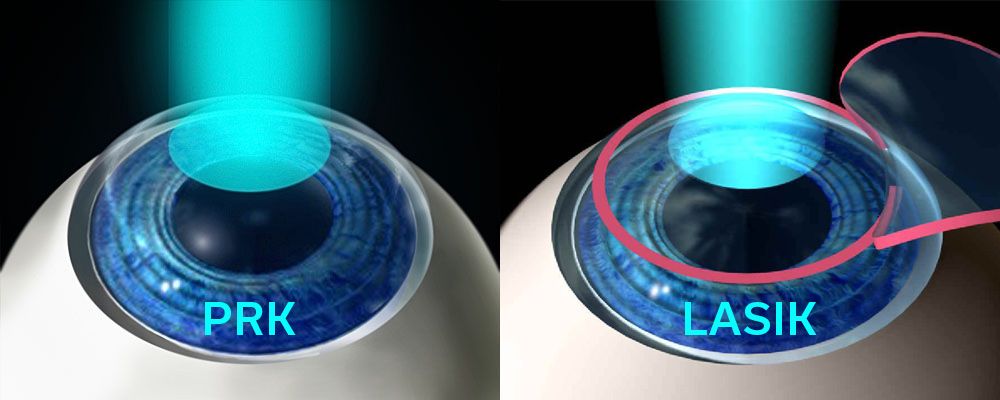
Comparison with Other Refractive Surgeries
LASIK: Faster visual recovery and less discomfort, but not suitable for patients with certain corneal conditions.
Standard PRK: Similar recovery profile but less customized compared to topography-guided PRK.
SMILE: A newer technique with a minimally invasive approach, but with less customization than topography-guided PRK.
Patients who undergo topography-guided PRK at the Khanna Vision Institute often experience a significant improvement in their visual acuity, with many reporting a reduction in the distortions and aberrations that were once a burden. The institute’s commitment to utilizing the latest advancements in corneal surgery, coupled with their team of highly skilled and experienced surgeons, makes them a trusted choice for individuals seeking effective treatment for keratoconus.